
Figure 2. AngII acutely regulates L‐type Ca current (I Ca) gating by oxidized PKARIα (protein kinase A type I‐α regulatory subunit).
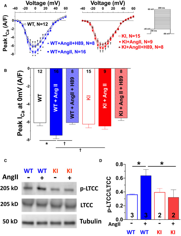
Mean data for peak I Ca voltage relationship (A) and peak I Ca at 0 mV (B) measured by whole‐cell patch‐clamp technique in isolated mouse ventricular myocytes (protocol depicted on the right in A). Acute exposure to AngII (angiotensin II; 1 μmol/L, 10 minutes) significantly enhances peak I Ca, which could be blocked by inhibition of protein kinase A (PKA) using the PKA inhibitor H89. This AngII‐PKA–dependent enhancement of I Ca was completely absent in knock‐in (KI) myocytes. Data are normally distributed (Shapiro‐Wilk test). *Indicates significance vs wild‐type (WT). †Indicates significance vs WT+AngII (2‐way repeated‐measures ANOVA, mixed‐effects model with Tukey post‐test). C and D, Original scans (C) and mean densitometric values (D) for Western blot analysis of L‐type Ca channel, alpha 1C subunit (CaV1.2) expression and serine 1928 phosphorylation (p‐LTCC [L‐type Ca channel]) in isolated hearts perfused with AngII (1 μmol/L, 10 minutes). Exposure to AngII significantly enhanced LTCC phosphorylation in WT but not KI mice. Data are normally distributed (Shapiro‐Wilk test). *Indicates significance vs WT+AngII using 1‐way ANOVA with Holm‐Sidak post‐test.