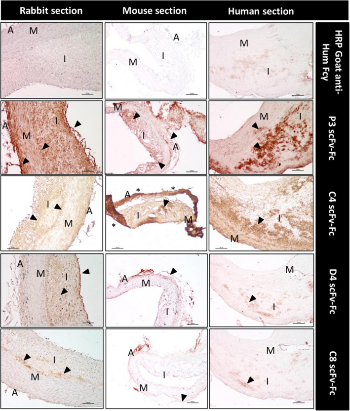
Figure 2. Immunohistochemical analysis of arterial tissue sections from atheromatous rabbits and Apoe−/− mice and human endarterectomy specimens using the indicated scFv‐Fc (single‐chain fragment variable fused to the crystallizable fragment of immunoglobulin G).

The different areas in transversal sections are identified: adventitia (A), media (M), and intima (I). Sections were incubated with the indicated scFv‐Fc antibodies (P3, C4, D4, and C8), followed by the HRP‐conjugated goat anti‐human Fcγ antibody, and the DAB substrate kit reagent. The yellow‐brown staining indicates the presence of the antigen recognized by the scFv‐Fc. No staining was observed in mouse and rabbit sections incubated with the secondary antibody alone, and there is low background noise in the human sections (upper panels). Scale bars, 100 µm. Nuclei were counterstained with hematoxylin. DAB indicates 3,3′‐diaminobenzidine; and HRP, horseradish peroxidase.
