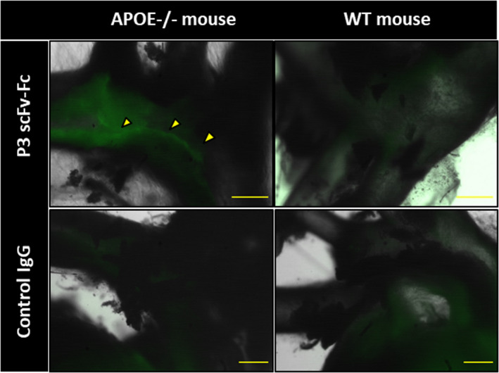
Figure 7. Ex vivo imaging of P3 scFv‐Fc (single‐chain fragment variable fused to the crystallizable fragment of immunoglobulin G) in Apoe−/− and wild‐type (WT) mice using a fluorescent ultramicroscope.

Fluorescence macroscopy analysis of P3 scFv‐Fc coupled to Alexa Fluor 568 dye after ex vivo injection in Apoe−/− and WT mice. A human immunoglobulin (Ig)G coupled to Alexa Fluor 568 was used as negative control antibody. P3 scFv‐Fc shows specific labeling of the atheroma in the Apoe− /− mouse (yellow arrowheads). Size bars: 500 μm.
