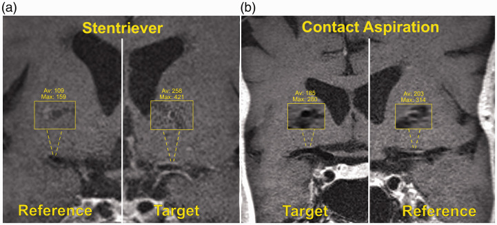
Figure 1.
(a) High-resolution magnetic resonance imaging (HR-MRI) coronal view of a patient who underwent mechanical thrombectomy (MT) with a stentriever. Inlets show the cross-sectional views of the reference (right middle cerebral artery (MCA)) and target (left MCA) vessels. Note the signal intensity (SI) difference between target and reference vessels (max/mean 421/258 vs. 159/109). (b) Coronal view of a patient who underwent MT with contact aspiration: inlets of the target (right MCA) and reference (left MCA) vessels show minimal SI difference (max/mean 280/185 vs. 314/203).

