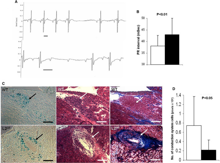
Figure 3. Conduction system physiology and histology of in‐frame LAMP2 gene exon 6 deletion (L2Δ6) hearts.

A, Representative traces recorded by telemetry in 15‐week‐old L2Δ6 mice: upper panel showed transient sinus node arrest and middle panel shoes transient atrioventricular block, Scale bars represent 50mSec. B, Average PR intervals (the time from the onset of the P wave to the start of the QRS complex on the electrocardiogram) of 15 weeks old WT (white bar) and L2Δ6 (black bar) mice, n=10/genotype, mean±SEM. C, Histology of the atrioventricular node and surrounding tissues in 15‐week‐old MnK/WT mouse (upper panels) and MnK/L2Δ6 mouse (lower panels). The hearts were stained with X‐Gal to identify β‐galactosidase activity in the conduction system cells (Blue dots in left panels), and then counterstained with hematoxylin–eosin (middle panels) and with Masson Trichrome (right panels). The atrioventricular node (arrow) in the MnK/ L2Δ6 hearts was more disorganized than MnK/WT hearts. Fibrosis and large vacuoles were detected in the atrioventricular region of MnK/L2Δ6 but not in the MnK/WT hearts. Scale bars represent 200μM. D, Quantitation of positive X‐Gal staining which represents conduction system cells. MnK/L2Δ6 hearts had less conduction cells (black bar) compared with MnK/WT (white bar). Analysis was done in consecutive section containing the atrioventricular node from 4 mice aged 15 to 20 weeks of each genotype, 3 sections per heart. Generalized Estimating Equation was used for statistical comparison. L2Δ6 indicates in‐frame LAMP2 gene exon 6 deletion; and WT, wild‐type.
