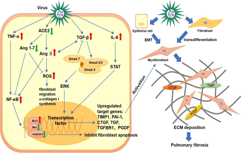
Fig. 2.
Signaling pathways of pulmonary fibrosis induced by the virus. Virus infection increases the expression of pro-fibrotic and pro-inflammatory cytokines such as TGF-β, TNF-α and IL-6 to promote pulmonary fibrosis. During this process, TGF-β/Smad, ERK and STAT pathways are activated and then upregulate profibrotic cytokines such as TIMP1, PAI-1, CTGF, TGF, TGFBR1, and PGDF. Besides, the decrease in ACE2 upregulates Ang II, leading to increased expression of NF-κB and ROS. NF-κB inhibits fibroblast apoptosis by upregulating Bcl-2 as well as downregulating Bax and caspase 3 to promote pulmonary fibrosis. ROS induces fibroblast migration and α-collagen I synthesis. Moreover, Ang II and TGF-β mutually reinforce each other. Eventually, a large number of myofibroblasts accumulate through EMT and fibroblast transdifferentiation, resulting in extracellular matrix deposition and pulmonary fibrosis

