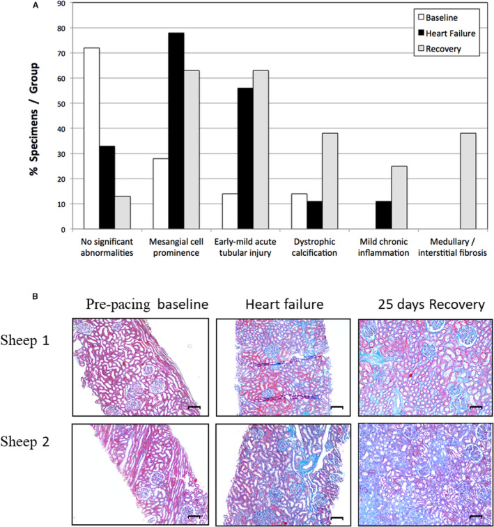
Figure 2. Serial histological responses in sheep during development of, and recovery from, acute decompensated heart failure.

A, Principal histological findings in serial kidney biopsies taken in 9 sheep before (baseline, n=7), after development of acute decompensated heart failure (induced by left ventricular pacing at 220 bpm for 14 days; n=9), and after 25 days of recovery following termination of pacing (recovery, n=8). (Note that mesangial cell prominence is not included here as a significant histologic abnormality.). B, Masson’s trichrome staining of representative serial kidney biopsies taken before (baseline) and after development of acute decompensated heart failure (induced by left ventricular pacing at 220 bpm for 14 days) and after 25 days of recovery following termination of pacing (recovery). Blue staining indicates collagen deposition. Scale bar=100 μmol/L.
