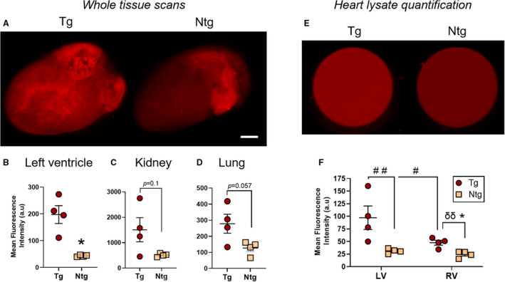
Figure 5. Collagen I‐targeted cyclic peptide enhancement of fibrotic mouse hearts and major organs in β2‐AR (β‐2‐adrenergic receptor) mice.

A through D, Representative ex vivo whole heart Odyssey images (A) and quantitation of cyclic peptide accumulation in the left ventricle (B), kidney (C), and lung (D) demonstrating cyclic peptide enhancement of perfused hearts and major organs in transgenic (Tg) vs nontransgenic (Ntg) β2‐AR mice 4 hours after peptide administration (0.5 mg/kg). E and F, Representative Odyssey scans (E) and quantification (F) of 96 wells containing left and right ventricular lysates from the hearts of these cyclic peptide–treated β2‐AR mice. Fluorescent signal enrichment in both chambers of the Tg mouse heart can be observed. Mean±SEM. N=4 per group. In (B through D and F), *P<0.05 by Mann‐Whitney test (1‐tailed). In (F), # P<0.05, ## P<0.01 by repeated‐measures 2‐way ANOVA with Sidak post hoc; δδ P<0.01 by an unpaired t test. Scale bar=1.8 mm. a.u. indicates arbitrary unit.
