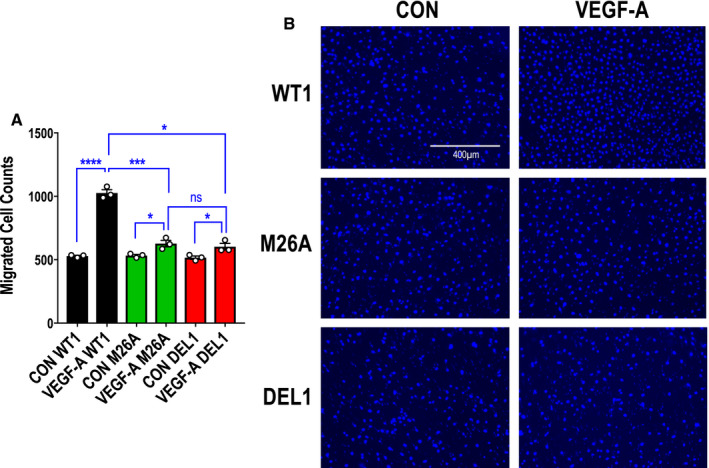
Figure 5. Effect of Ser26 mutation and Egr‐1 deletion on endothelial migration.

A, Migration assays were performed with CRISPR/Cas9 clones WT1, M26A, and DEL1 using a dual‐chamber model containing VEGF‐A165 (vascular endothelial growth factor‐A) (25 ng/mL) in the lower chamber. Data represent the means of the means of 3 biologically independent experiments±SEM. Significance was assessed by Mann‐Whitney or t test. *P≤0.05; ***P<0.001; ****P<0.0001. B, Representative DAPI‐stained nuclei in each condition. The cells were photographed under 10× magnification. CON indicates control cells (in 0.1% FBS) in the model without VEGF‐A165; DEL, deletion; Egr‐1, early growth response‐1; M, mutant; ns, not significant; and WT, wild‐type.
