
Figure 3. Obese rats exhibited an increase in pericardial adipose tissue (PAT)–derived leptin and enhanced cardiac oxidative stress accompanied by a decrease in Na+/K+‐ATPase (NKA) activity.
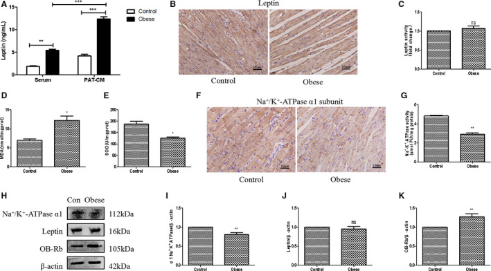
A, ELISA analysis of leptin levels in both serum and PAT conditioned medium (PAT‐CM) of control and obese rats. B and C, Immunohistochemical (IHC) analysis of cardiac leptin expression of control and obese rats. D, Detection of cardiac malonaldehyde (MDA) assay of control and obese rats. E, Detection of cardiac superoxide dismutase (SOD) assay of rats in 2 groups. F and G, IHC analysis of cardiac NKA α1 subunit expression of control and obese rats. H through K, Western blot analysis of cardiac α1 subunit of NKA, leptin, and leptin receptor (OB‐Rb) of control and obese rats. Two‐way ANOVA was used for A; t‐test was used for D, E, and I through K; and Mann‐Whitney U test was used for C and G. Data are presented as mean±SEM. For D, E, and I through K, n=3/group; for A, C, and G, n=5/group. Ns indicates insignificant. *P<0.05 vs control, **P<0.01 vs control, and ***P<0.001 vs control.