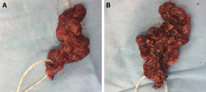
Figure 1.
A 7-cm polypoid tubulovillous adenoma extending from the ampulla of Vater down to D3, removed by means of open surgical ampullectomy-excision of adenoma en block, following cholecystectomy and catheterization of the ampulla for identification. Preoperative biopsies showed low-grade dysplasia (LGD) and this 74-year old patient with severe comorbidities was initially counselled for pancreatoduodenectomy. Meticulous preoperative endoscopic evaluation revealed its polypoid configuration, possibly emanating from a mucosal stalk, which was confirmed intraoperatively. A: The ampulla is encircled by a fine catheter. Final histology confirmed the presence of a tubulovillous adenoma with extensive LGD and focal high-grade dysplasia. All margins were clear of tumour or dysplasia. (V. Mavroeidis’ archive). B: Inferior aspect of the specimen, depicting the duodenal margin along the tumour, and the insertion point of the catheter into the ampulla. (V. Mavroeidis’ archive).

