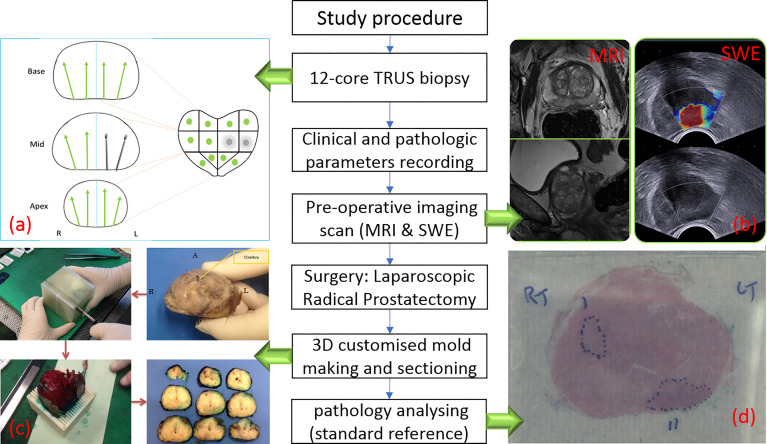
Figure 1.
Flow chart of study procedure. (A) TRUS biopsy result with two positive biopsy cores. (B) One suspicious lesion in peripheral zone is shown in MRI (left) and SWE (right) images. (C) Post-prostatectomy specimen sectioning in steps (23). (D) Histopathology photo after analysing.

