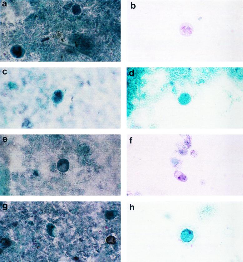
FIG. 1.
Composite photograph showing the appearances of Entomeba species and I. bütschlii in slides prepared by each of the four fixation and staining methods under study. (a) E. histolytica, PVA. (b) E. histolytica, PF. (c) E. histolytica, EC. (d) E. coli, PS. (e) I. bütschlii, PVA. (f) I. bütschlii, PF. (g) I. bütschlii, EC. (h) I. bütschlii, PS. Magnification, ×750.

