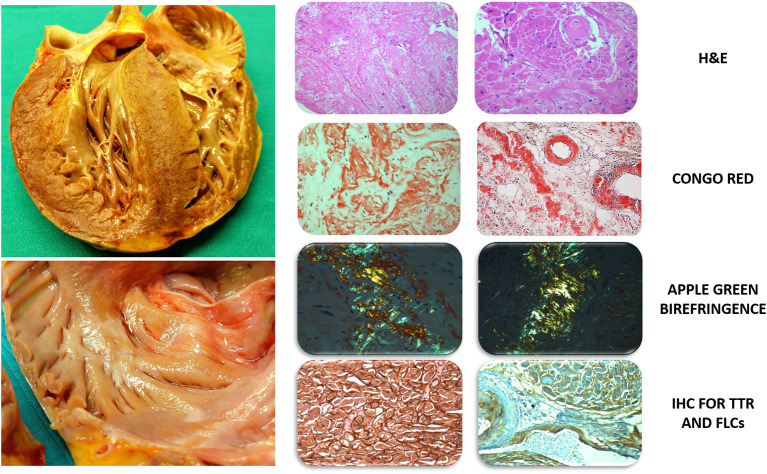
Figure 1.
Gross evaluation of unfixed heart and histological characterization with dedicated staining. Left Top: massive cardiac hypertrophy of the left ventricle involving the subvalvular aortic region extending to the right ventricle, the papillary muscles and the inter-atrial septum. The heart exhibits a heterogeneous color with pale areas and faint yellowish tone. Bottom: increased atrial wall thickness with diffuse granulations on the endocardial surface and nodular deposits of amyloid. Right: left column (optical field 10×) and right column (optical field 20×): H&E, severe amyloid infiltration in the interstitium and vessels with degeneration of myocytes; Congo Red and polarized light microscope, Congo Red binding to amyloid fibrils showing the typical apple-green birefringence and revealing predominant interstitial infiltration; amyloid infiltration involves the interstitial space and vessel walls, including the sinoatrial nodal artery; IHC for TTR and FLCs, positivity of transthyretin (left, 40×) and immunoglobulin light chain Kappa (right, 20×) in advanced cases of cardiac amyloidosis. H&E, Hematoxylin and Eosin; FLCs, Free Light Chains; IHC, Immunohistochemistry; TTR, Transthyretin.

