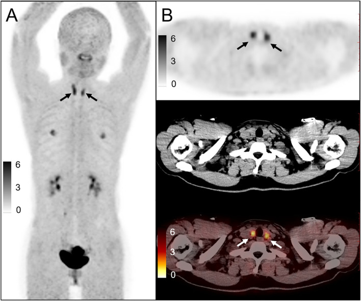
Figure 1.
A 32-year-old woman underwent 68Ga-FAPI PET/CT for rectal cancer after surgery [(A): maximum intensity projection image; (B): upper, PET, middle, CT, lower, PET/CT image]. The uptake of thyroid 68Ga-FAPI increased diffusedly, and the SUVmax was about 4.6, and TBR was 7.54. CT showed a decrease in density. Ultrasound examination showed that the echo of the thyroid gland was reduced and the blood flow was uneven. A fine needle aspiration biopsy of the thyroid revealed chronic thyroiditis. Thyroid function test revealed abnormal TSH and thyroid antibody levels (TSH: 16.117, reference range, 0.38–5.57 mIU/L; FT3:2.07, reference range, 1.80–3.80 pg/ml; FT4: 0.86, reference range, 0.78–1.86 ng/dl; TPOAb: 872.18, reference range, 0.0–2.6 IU/ml; TgAb: 620.47, reference range, 0.0–14.58 IU/ml), conforming the diagnosis of lymphocytic thyroiditis.

