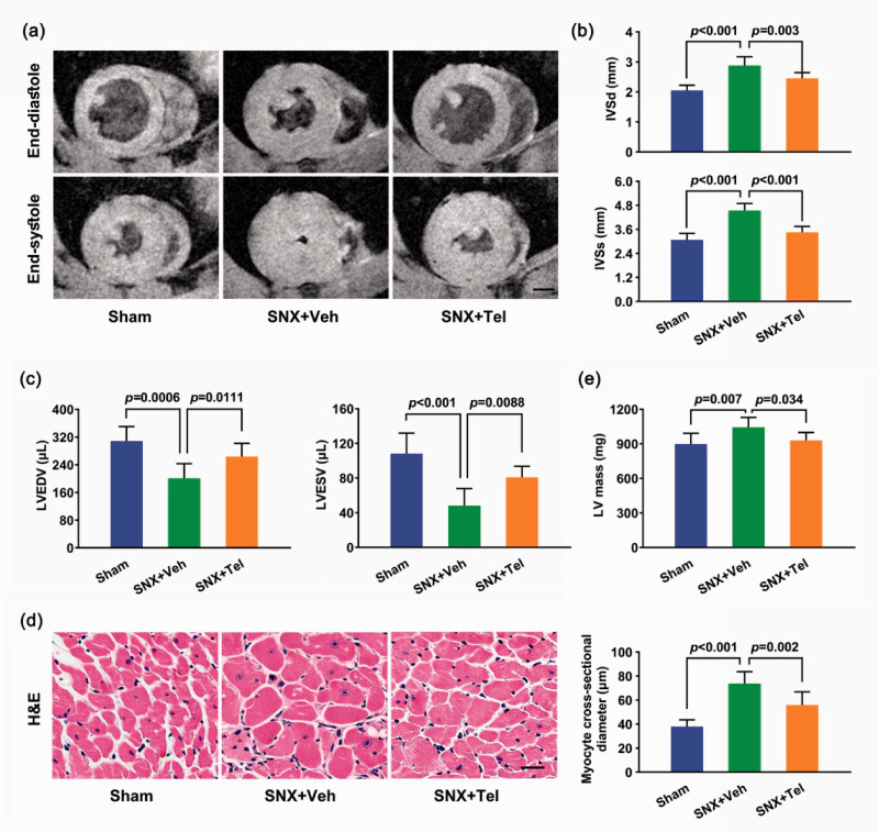
Figure 4.
Left ventricular hypertrophy measurement by cardiac MRI and histology. (a) Cardiac Cine MRI of the same slice of the heart at end-diastole (top row) and end-systole (bottom row) of the three groups at week 16 and (b) the mean ± SD of IVSd and IVSs as well as (c) the mean ± SD of LVEDV and LVESV at week 16 after the surgery. (d) Cardiac H&E staining of myocytes in cross-section at week 16 and the calculated mean ± SD of myocyte cross-sectional diameter. (e) The mean ± SD of LV mass at week 16. Scale bar in (a), 2.5 mm. Scale bar in (d), 50 μm. MRI indicates magnetic resonance imaging; IVSd: interventricular septal thickness at end-diastole; IVSs: interventricular septal thickness at end-systole; LVEDV: left ventricular end-diastolic volume; LVESV: left ventricular end-systolic volume; H&E: hematoxylin & eosin; LV: left ventricular. (A color version of this figure is available in the online journal.)

