Figure 1.
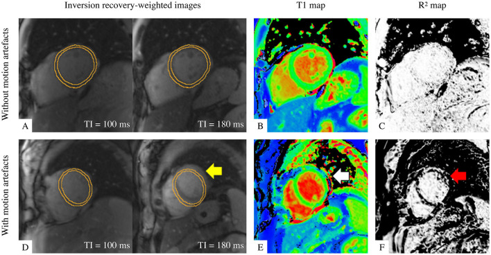
Illustration of T1 maps with good quality (top row) and with motion artefact (bottom row). (A,D) Two examples out of seven of inversion-recovery weighted (IRW) images required for T1 map reconstruction are shown, time-stamped with their corresponding inversion times (TI) and overlaid by identical manual myocardial contours for identifying motion. (B,E) ShMOLLI T1 maps. (C,F) R2 quality control maps. A good quality T1 map is indicated by (A) myocardium in same position and (C) “all white” in the left ventricular myocardium indicating high T1 fitting confidence. A T1 map with motion artefact is evident by misalignment in IRW images (yellow arrow), suspicious features in T1 map (white arrow) and dark bands in R2 map in the left ventricular myocardium as evidence of poor T1 fitting (red arrow).

