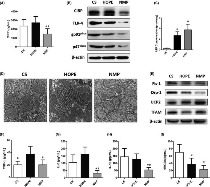
FIGURE 4.

NMP reduces CIRP secretion and mitochondrial dysfunction in the DCD liver. The DCD livers were preserved for 4 h in vitro using CS, HOPE and NMP, respectively (n = 6). (A) The expression of CIRP in the perfusate of the three different groups. (B) The expression of CIRP, TLR‐4, and NADPH oxidase in liver tissues. (C) ATP content in the livers of the three groups. (D) TEM images of liver tissues. Liver tissues in the NMP group had a high mitochondrial density, clear mitochondrial membrane and cristae, and without expanded endoplasmic reticulum (magnification 20000×). (E) Western blot analysis of mitochondrial proteins. The expression of inflammatory factors including TNF‐α (F), IL‐6 (G), IL‐1β (H), and HMGB1 (I) in the perfusates. *p < 0.05 versus the CS group, # p < 0.05 versus the HOPE group. Scale bars: 1 μm (D)
