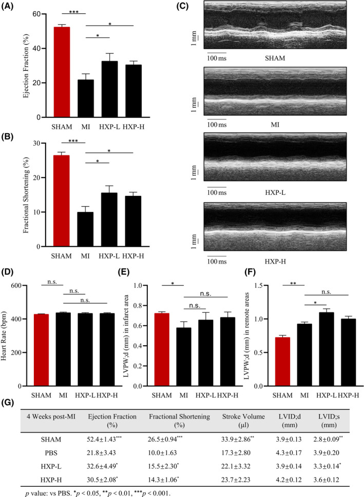
FIGURE 1.

HXP improves cardiac function following ischaemic injury. (A‐B) Ejection fraction (EF%) and fractional shortening (FS%) parameters in mice administrated with PBS, low‐dose HXP‐L (3 mg kg d−1) or high‐dose HXP‐H (9 mg kg d−1) at 4 weeks post‐MI. n = 5 or more in each group, * p < 0.05, ** p < 0.01, *** p < 0.001. (C) Representative M‐mode echocardiographic images showing LV end‐systolic and end‐diastolic dimensions in mice administrated with PBS, low‐dose HXP‐L (3 mg kg d−1) or high‐dose HXP‐H (9 mg kg d−1) at 4 weeks post‐MI. (D‐G) Echocardiographic measurements of heart rate (D), LV end‐diastolic wall thickness in diastole at the infarct border zone (E), and remote zone (F), and cardiac function parameters (G) in sham, PBS, low‐dose HXP‐L (3 mg kg d−1), or high‐dose HXP‐H (9 mg kg d−1) mice at 4 weeks post‐MI. Data are presented as mean ± sem. * p < 0.05, ** p < 0.01, *** p < 0.001, n.s., no significance. n = 5 or more in each group. LVID;d—Left Ventricular Internal Dimension; diastolic; LVID;s—Left Ventricular Internal Dimension; systolic
