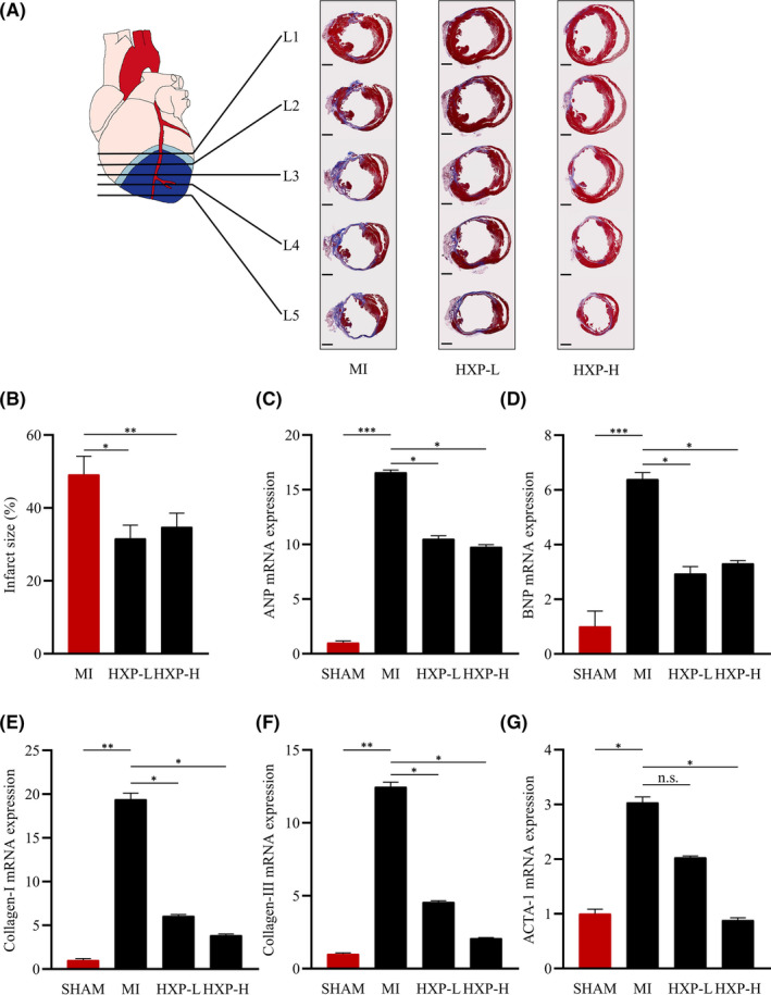
FIGURE 2.

HXP prevents MI‐induced cardiac remodelling. (A) Masson's trichrome staining of hearts at 4 weeks post‐MI in mice administrated with PBS, low‐dose HXP‐L (3 mg kg d−1), or high‐dose HXP‐H (9 mg kg d−1) showing 5 different levels (L1‐L5) of heart transverse cross sections from the site of ligation towards the apex. Muscle fibres are stained red, and collagen‐rich fibrotic regions are stained blue. n = 9 or more for each group. Scale bar, 1 mm. (B) Quantification of infarct size in cardiac sections. Infarct size (%) = Mean endocardial infarct arc lengths at L3‐L4 divided by mean left ventricular endocardial arc lengths at L3‐L4 × 100. (C‐G) Relative mRNA expression levels of cardiac fibrosis genes (collagen‐I and collagen‐III) and heart failure genes (ANP, BNP, skeletal α‐actin) in the infarct regions of mice treatment with PBS, low‐dose HXP‐L (3 mg kg d−1) or high‐dose HXP‐H (9 mg kg d−1) at 2 weeks post‐MI. Data are presented as mean ± sem, n.s., no significance, * p < 0.05, ** p < 0.01, *** p < 0.01. n = 5 or more for each group
