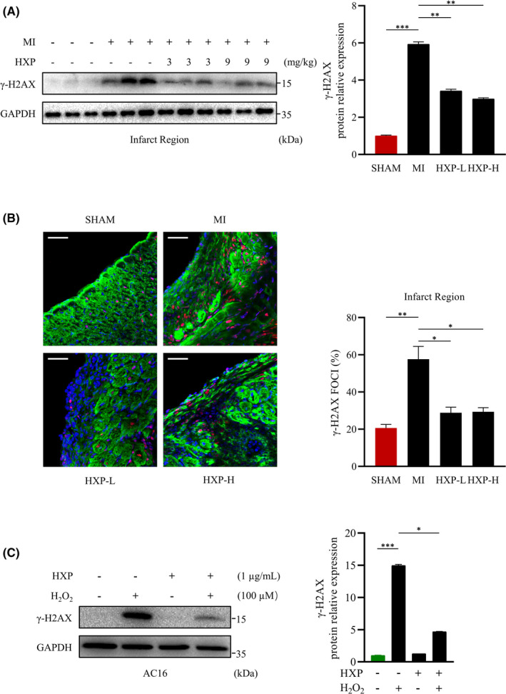
FIGURE 3.

HXP attenuates ischaemia‐induced DNA damage in vitro and in vivo. (A) Representative immunoblots (left) and quantification (right) showing γ‐H2AX expression in ischaemic hearts from mice treated with PBS, low‐dose HXP‐L (3 mg kg d−1) or high‐dose HXP‐H (9 mg kg d−1) at 1‐week post‐MI. GAPDH was used as the loading control. Data are presented as mean ± sem, ** p < 0.01, *** p < 0.001. n = 3 for each group. (B) Representative immunofluorescence staining (left) and quantification (right) of γ‐H2AX foci (red), cardiac muscle marker Troponin T (green) and DAPI (blue) in the infarcted LV myocardium in SHAM‐operated mice and those treated with PBS, low‐dose HXP‐L (3 mg kg d−1) or high‐dose HXP‐H (9 mg kg d−1) at 1‐week post‐MI. Data are presented as mean ± sem, * p < 0.05, ** p < 0.01. n = 4 for each group. Scale bar, 50 μm. (C) Representative immunoblot (left) and quantification (right) showing expression of γ‐H2AX following pretreatment with 1 µg ml HXP for 2 hours and treatment with 100 µM H2O2 for 30 minutes in AC16 cardiomyocytes. GAPDH was used as the loading control. Data are presented as mean ± sem, * p < 0.05, *** p < 0.001 versus control group. n = 3 for each group
