FIGURE 5.

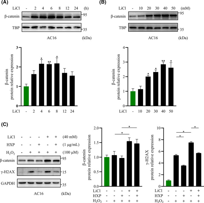
HXP attenuates H2O2‐induced DNA damage via inhibition of Wnt/β‐catenin pathway. (A) Representative immunoblot (top) and quantification (bottom) showing the expression of nuclear β‐catenin following LiCl stimulation at different time intervals in AC16 cardiomyocytes. TBP was used as the loading control. n.s., no significance, * p < 0.05, ** p < 0.01 versus control group. n = 3 for each experiment. (B) Representative immunoblot (top) and quantification (bottom) showing the expression of nuclear β‐catenin following LiCl stimulation at different concentrations in AC16 cardiomyocytes. TBP was used as the loading control. n.s., no significance, * p < 0.05, ** p < 0.01 versus control group. n = 3 for each experiment. (C) Representative immunoblot (left) and quantification (right) showing the expression of γ‐H2AX and β‐catenin following pretreatment with 1 µg ml−1 HXP for 2 hours, 40 mM LiCl for 4 hours, and subsequent treatment with 100 µM H2O2 for 30 minutes. GAPDH was used as the loading control. Data are presented as mean ± sem, * p < 0.05. n = 3 for each
