
FIGURE 6.
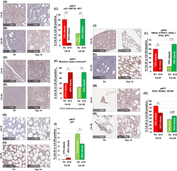
CA IX and/or CA XII is expressed in non‐FLT3 mutated AML patients failing induction chemotherapy. CA IX and XII staining of BM samples from a diverse panel of non‐FLT3 mutated AML patients who had residual disease on their day 14 marrow assessment (A–O). Consistent with data obtained from AML xenograft studies, four out of five patients showed significantly increased CA IX and/or CA XII staining in leukemic blasts remaining after induction chemotherapy. All patients were treated with the “7 + 3” induction regimen. Results shown represent the mean ± SEM % CA IX or XII positivity. Statistically significant changes in the percentage of CA staining are indicated (* <0.05; **p < 0.01; ***p < 0.001). Leukemic blast cell percentages were quantified per clinical flow cytometric immunophenotyping by the Indiana University Health Pathology Laboratory. Corresponding H&E stained sections from the BM are shown in Figure S4A–H