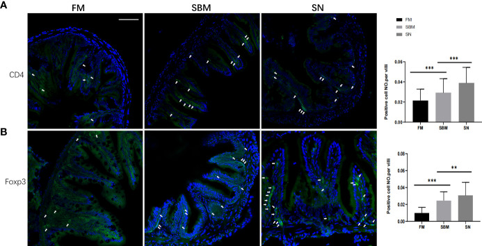
Figure 2.
Lymphocyte-related intestinal protein expression reflected by immunofluorescent signals. CD4+ (A) or Foxp3+ (B) immunofluorescence signals (green) in adult fish fed with FM, SBM, and SN diets. The quantification of the ratio, which was calculated by dividing positive cell number with intestinal villi’s length, was shown beside the typical images (at the right side). Typical signals are indicated by arrows. The blue signals represented DAPI-stained cell nucleus. FM, fish meal diet; SBM, soybean meal diet; SN, sinomenine supplementary SBM diet. Scale bar: 100 μm. ** represented p < 0.01, and *** represented p < 0.001.

