Fig. 2.
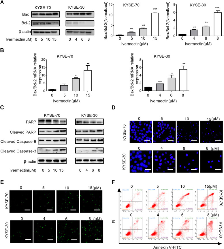
Ivermectin promotes apoptosis of ESCC cells. Western blotting (A) and qRT-PCR (B) analysis for the expression levels of Bax and Bcl-2 after ivermectin treatment; C. The expression of caspase cascade (cleaved-caspase 9, cleaved-caspase 3, PARP, cleaved PARP) were analyzed by western blotting; D. Cells were observed by fluorescence microscopy after staining with Hoechst 33342, Scale bar = 100 μm; E. Ivermectin-induced ESCC cells apoptosis was determined by TUNEL assay (200×), Scale bar = 50 μm; F. Ivermectin-induced ESCC cells apoptosis was determined by PI/Annexin-V assay using flow cytometry. *P < 0.05, **P < 0.01; ***P < 0.001.

