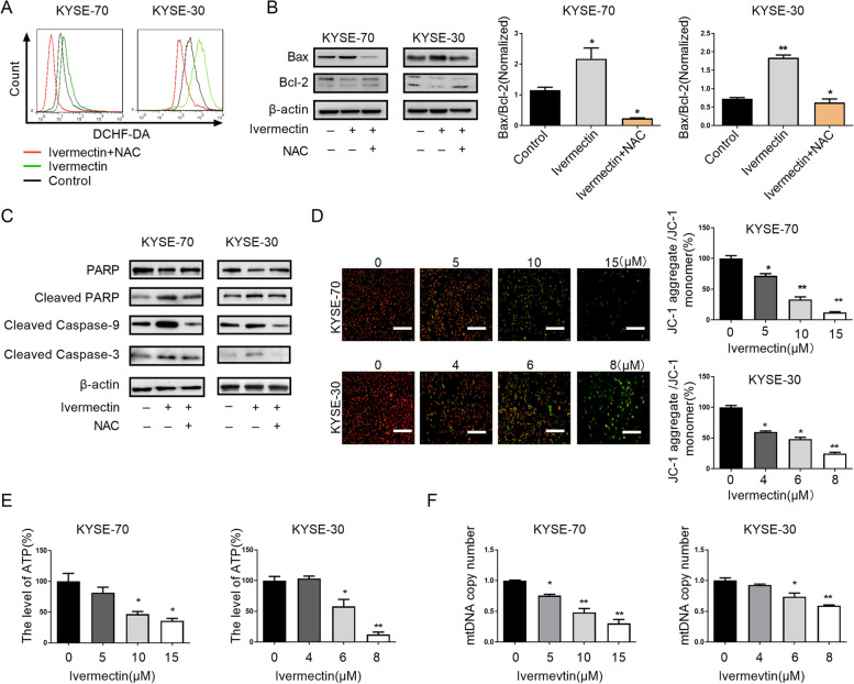
Fig. 3.
Ivermectin mediates mitochondrial dysfunction of ESCC cells. A. Intracellular ROS levels were investigated by DCFH-DA fluorescence in ESCC cells treatment with indicated; B. The expression levels of Bax and Bcl-2 were determined by western blot analysis; C. The expression of caspase cascade (cleaved-caspase 9, cleaved-caspase 3, PARP, cleaved PARP) were analyzed by western blotting; D. Mitochondrial membrane potential was observed by fluorescence microscope at 200 × magnifications, Scale bar = 50 μm; E. ATP production of ESCC cells after treated with ivermectin was detected; F. The mitochondrial DNA copy number in ESCC cells treatment with invermectin was evaluated using qRT-PCR. *P < 0.05, **P < 0.01.

