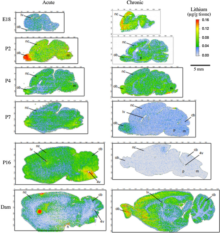
Fig. 7.
Distribution of Lithium in the brain following acute or long-term exposure to lithium at different ages. Representative sagittal sections of brains at different ages from animals exposed to lithium acutely (left hand panels) and long-term (right hand panels). Olfactory bulbs are to the left in all sections. The dimensions of the brain sections are approximately to scale but due to dehydration in preparation of tissue they can vary between ages. At most postnatal ages there is a concentration of lithium in the olfactory bulbs and more generalised lower-level distribution in the rest of the brain. There was a similar distribution in the postnatal brains of animals treated long-term, but with lower intensity. Control sections were always blank. More sections of treated and control brains are in Additional file 3: Fig S2. P0 is not included in the long-term treated group (right hand panels) as in long-term treated pups the dam’s treatment started from the day of birth. Labels: 4v (4th ventricle), cb (cerebellum), h (hypothalamus), lv (lateral ventricle, m (medulla), nc (neocortex) ob (olfactory bulb), p (pons)

