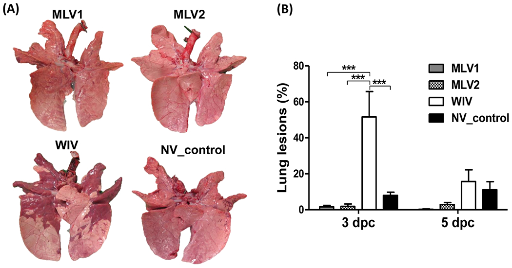
Fig 2. Macroscopic lung lesions in pigs after challenge.

(A) Ventral surfaces of lungs from representative pigs in each group at 3 dpc are shown. (B) Macroscopic lung lesions of challenged pigs are presented as the average percentage ± SEM of gross lesions of three pigs in each group at 3 and 5 dpc. The asterisks (*) represent a statistically significant difference between groups (***: p<0.001).
