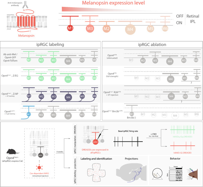
Fig. 3.
Diverse toolkit for studying ipRGCs circuits. Top, differential melanopsin expression in M1–M6 ipRGCs. Anti-melanopsin antibody binds to the N-terminus extreme, labeling M1–M3 ipRGC subtypes. ipRGC labeling box: Opn4-GFP line labels M1–M3 ipRGCs in the adult mouse retina and also M4 cells prior to P14 [43]. The Opn4-tdTomato line labels similar subtypes to Opn4-GFP. Opn4Cre/+; Z/EG labels M1–M6 ipRGCs [7]. Opn4Cre/+; Z/AP line labels somas and axons of M1–M6 cells after AP staining [7]. Opn4LacZ/+ line labels M1 ipRGCs somas and axons after X-gal staining [2]. ipRGC ablation box: in the Opn4aDTA line M1 ipRGCs are ablated [19]; in Opn4DTA, M1–M3 cells are likely ablated and Opn4Cre/+; R26iDTR mice potentially all ipRGCs are ablated [49, 53]. In Opn4Cre/+; Brn3bZ-DTA, all ipRGCs are ablated except Brn3b-negative M1ipRGCs [99]. Bottom, use of adeno-associated viruses (AAV) to manipulate ipRGC activity through DREADDs (top) or to label somata and axons of ipRGCs (bottom) [50–52, 55–60]

