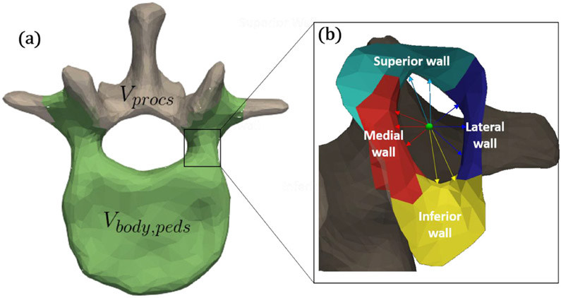
Figure 3.
Illustration of components defined within a vertebral level. (a) Vertices belonging to the vertebral body and pedicle distinguished from those of the spinous process (L1 SSM is shown). (b) Sub-components within the pedicle corridor: medial, lateral, superior, and inferior walls (L1 left pedicle is shown). Also shown in (B) are pertinent distances from the centerline (green): medial (dmed,i,ped), lateral (dlat,i,ped), superior (dsup,i,ped), and inferior (dinf,i,ped).

