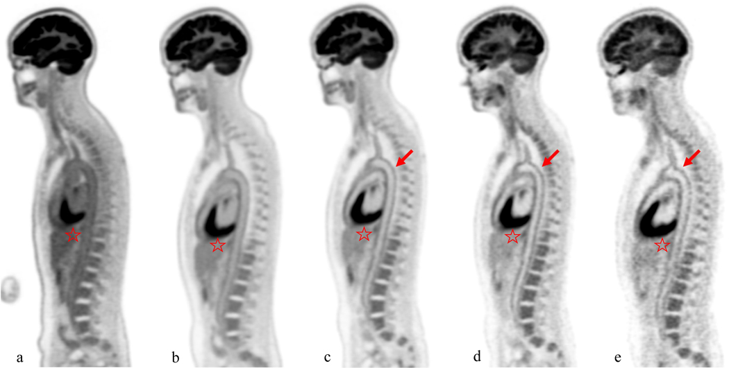
Figure 1:
Female, 41 year-old, 53 kg healthy volunteer subject. Sagittal slice images at the level of the aortic arch were obtained at 40 minutes (a), 90min (b), 3 h (c), 6 h (d), and 9 h (e) after injection of 10mCi of 18F-FDG. The aorta demonstrates the decreasing blood pool activity over time, leading to progressively higher signal to noise at the level of the aortic wall (arrows) on delayed images. Also brain and liver uptake (star) decreases on delayed images. However, the noise increases on delayed images leading to image quality at 9 hours comparable to image quality of a conventional PET/CT scan obtained at 60 minute.

