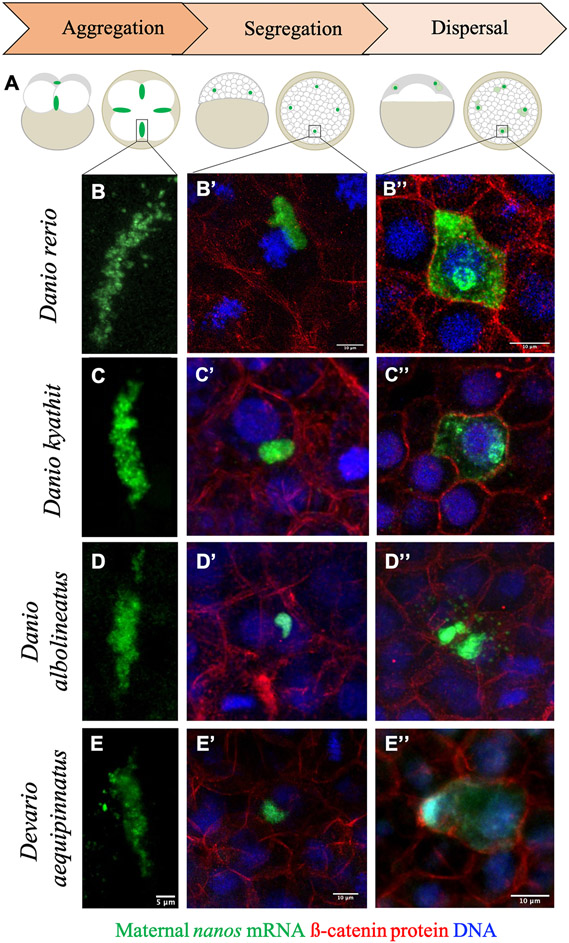
Figure 3.
Key stages of germ plasm localization dynamics across Danionin species. A) Diagram depicting an overview of germ plasm localization during early embryogenesis in zebrafish. Germ plasm ribonucleoparticles (green) in zebrafish form extended aggregates at the distal end of the first two embryonic cleavage furrows (1 hpf; aggregation; left), which become cellularized and segregate asymmetrically during cell divisions throughout the late cleavage through mid-blastula period of development (~1.5 hpf – 4 hpf; asymmetric segregation; middle), before dispersing to fill the cytoplasm of PGCs beginning at the dome stage through early gastrulation (~4.3 hpf – 6 hpf; dispersal; right). B-B”) Micrographs of germ plasm localization and morphology in zebrafish (Danio rerio) embryos during each of the highlighted stages, represented by the germ plasm marker nanos RNA (green) visualized via fluorescence in situ hybridization and confocal microscopy. B’ and B’’ also depict cell boundaries with beta catenin immunolabeling (red) and nuclei with DAPI stain (blue). The general appearance and localization patterns of germ plasm at these stages are consistent across species, including Danio kyathit (C-C”), Danio albolineatus (D-D”), and Devario aequipinnatus (E-E”).

