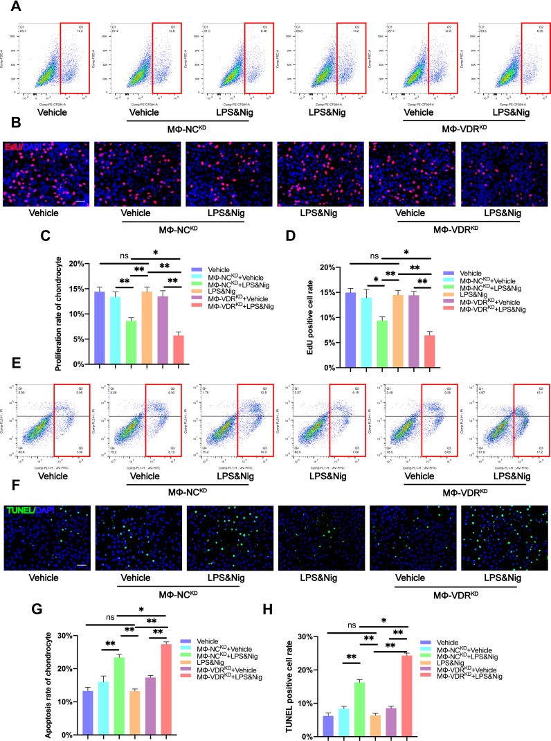
Figure 5.
Macrophage VDR partly reverses decrease in chondrocyte proliferation rate and increase in chondrocyte apoptosis rate caused by NLRP3 activation. (A) Proliferation rate of chondrocytes was determined by flow cytometry analysis after EdU assay. (B) Proliferation rate of chondrocyte as determined by immunofluorescence. Scale bar = 200μm. (C) Flow cytometry analysis of results from EdU assay (n = 3, one-way ANOVA). (D) EdU positive cell rate of each group (n = 3, one-way ANOVA). (E) TUNEL assay for apoptosis rate of chondrocytes determined by flow cytometry analysis. (F) Apoptosis rate of chondrocytes as determined by immunofluorescence. Scale bar = 200μm. (G) Flow cytometry analysis of TUNEL assay results (n = 3, one-way ANOVA). (H) TUNEL positive cell rate for each group (n = 3, one-way ANOVA). Data are presented as mean ± SD. ns, no significance; *p <0.05; **p <0.01.

