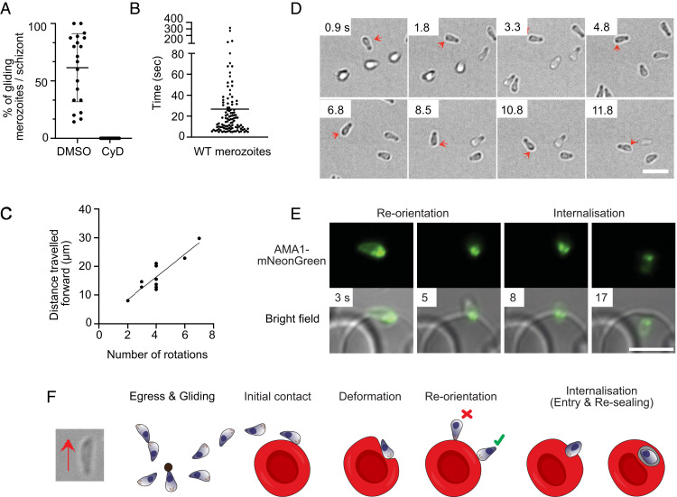
Fig. 2.
Gliding motility of P. knowlesi merozoites. (A) The percentage of merozoites within a P. knowlesi schizont which exhibit motility, for both DMSO-treated parasites (mean = 62.5%) and 0.1 μM cytochalasin D (CyD; IC50 = 0.023 ± 6.7 nM)-treated parasites (no gliding observed). A “motile” merozoite was defined as having demonstrated directional forward motion along the surface of the coverslip for at least five continuous seconds. Each dot is representative of one schizont (n = 20). Error bars denote ±1 SD. (B) The total time each motile P. knowlesi merozoite (n = 109; median = 15 s) spent gliding during the 10-min imaging window post egress. Error bars indicate interquartile range. (C) Number of rotations that merozoites completed plotted against the distance traveled for each glide (n = 10). As the number of rotations increased, so did the distance traveled forward, indicating rotation drives forward motion (Pearson correlation coefficient, R = 0.88). (D) Time-lapse imaging demonstrating a P. knowlesi merozoite rotating as it glides. Red arrows indicate a dark spot located to one side of the wider end of the merozoite, which shifts to the opposite side (shown in subsequent frames), as it turns, and then back to the original position to complete a full rotation (Movie S6). (Scale bar, 5 μm.) (E) Time lapse imaging depicting a P. knowlesi merozoite with mNeonGreen-tagged AMA1 invading an erythrocyte. Left and Left Center demonstrate reorientation of the wider end of the merozoite to align with the erythrocyte membrane. This is followed by (Right Center) the formation of the moving junction, depicted as two green dots at the merozoite-erythrocyte interface, and, finally, (Right) entry into the host cell. (Scale bar, 5 μm.) (F) Schematic illustrating gliding and erythrocyte invasion. Gliding proceeds with the wider, apical end of the merozoite leading. During gliding, merozoites stretch, and a pointed protrusion can be seen at the wider end of the zoite (left-hand bright-field image), which engages with the erythrocyte membrane upon reorientation and internalization. Reorientation of the zoite to make a contact of wider end (green tick), and not the thinner end of the zoite as previously hypothesized (red cross), with the erythrocyte membrane occurs prior to entry. During internalization, constriction of the apical end of the zoite causes the basal end to expand. Finally, after entry is complete, the parasite resides in a parasitophorous vacuole where its development continues.

