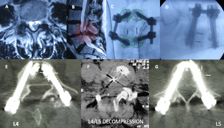
Figure 5.

An illustrative transforaminal lumbar interbody fusion (TLIF) case shows axial (A) and sagittal (B) magnetic resonance imaging scans demonstrating a huge right synovial cyst at the L4-L5 level. Anterior-posterior (C) and lateral (D) intraoperative fluoroscopic views illustrating TLIF and transpedicular screw fixation augmented with bone cement because of osteopenia evident on the preoperative bone density scan. The postoperative axial computed tomography scan images (E–G) validate the L4-level cyst resection, adequate position of the interbody fusion cage, and the L4 and L5 pedicle screws without breaches.
