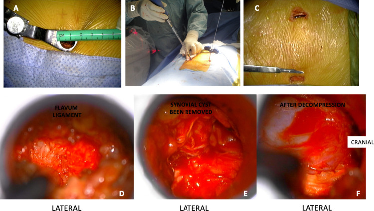
Figure 6.

Continuation of the same illustrative case shown in Figure 5. A 14-mm tube was placed for right-sided transforaminal lumbar interbody fusion and synovial cyst resection (A). Bilateral transpedicular screw fixation after transforaminal interbody fusion and cyst resection was performed (B). Bilateral skin incisions (right side at the bottom, left side at the top) were made (C). The ligamentum flavum was identified (D), and the synovial cyst (at the top) was noted to be adherent to it (E). The decompressed dural sac was visualized after completion of the cyst resection (F).
