Figure 5.
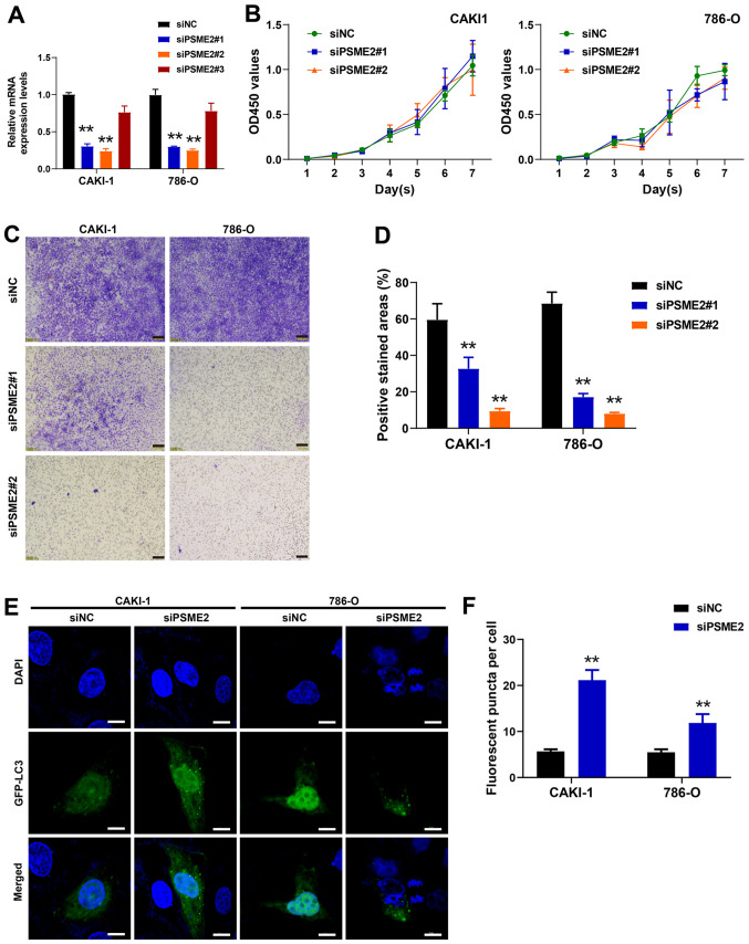
PSME2 promotes renal cancer cell invasion and inhibits autophagy. (A) mRNA expression levels of PSME2 were examined using reverse transcription-quantitative PCR. **P<0.01. (B) Cell proliferation of the CAKI-1 and 786-O cell lines transfected with siNC and siRNA was analyzed using a Cell Counting Kit-8 assay. (C) Transwell invasion assay was used to detect cell invasion following transfection with siRNA targeting PSME2. Scale bar, 100 µm. (D) The percent of positive stained areas in invaded cells per field. **P<0.01. (E) Knockdown of PSME2 induces autophagy in the CAKI-1 and 786-O cell lines using confocal fluorescence microscopy. The cells were transfected with siPSME2#2 and GFP-LC3 for 24 h. Nuclei were labelled with DAPI (blue). Scale bar, 10 µm. (F) The fluorescent puncta per cell were quantified. **P<0.01. PSME2, proteasome activator complex subunit 2; si, small inhibiting; NC, negative control; GFP, green fluorescent protein.

