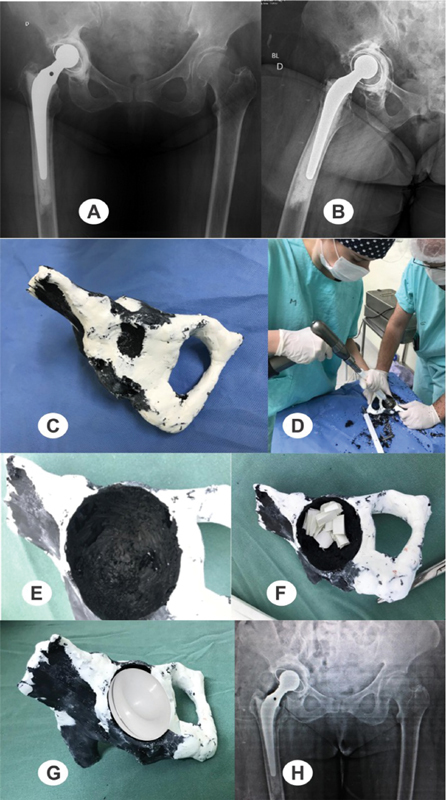
Fig. 1.

Preoperative radiographs of the pelvis ( A ) and lateral view of the right hip ( B ) at the time of the review, revealing an important acetabular bone failure with loosening of the components. ( C ) View of the printed prototyping model; ( D ) residents training with prototyping models; ( E ) cavity and ( F ) acetabulum preparation with visualization of the bone defect and its filling with chopped, impacted bone; ( G ) planning of the size and position of the implant; ( H ) postoperative radiograph of the revision of the right total hip arthroplasty.
