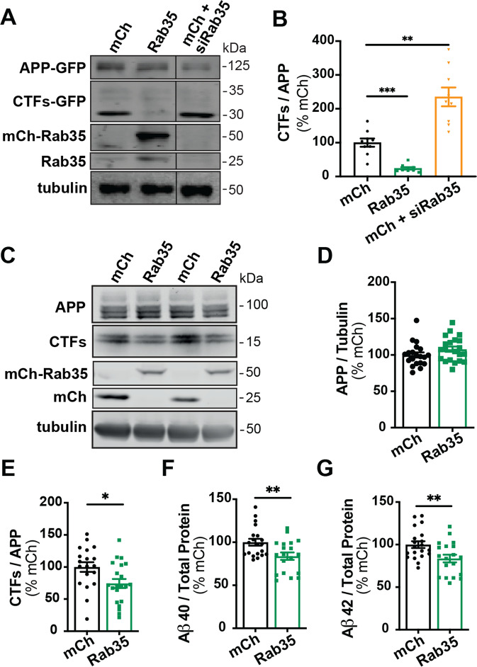
Fig. 2. Rab35 expression suppresses APP processing.
A, B Representative immunoblots and quantification of APP CTFs from N2a cells co-expressing APP-GFP with mCherry, mCh-Rab35, or mCh+siRNAs against Rab35 (siRab35). Immunoblots were probed for GFP, tubulin, and Rab35, with values normalized to tubulin and expressed as % of mCh control (***PmChCtrl vs. mCh-Rab35 = 0.0006, **PmChCtrl vs. mCh + siRab35 = 0.0029; one-way ANOVA with Welch’s correction, n = 9 samples/condition, 1 experiment). C–E Representative immunoblots and quantification of APP CTFs from iPSC-derived cortical neurons expressing mCh or mCh-Rab35. Blots were probed for APP, tubulin, and mCherry, with values normalized to tubulin and expressed as % of mCh control (*P = 0.017; unpaired t-test, n = 19–21/condition, four independent cultures). F, G Measurement of Aβ peptides secreted by iPSC-derived human neurons transduced with mCherry or mCh-Rab35. Values are normalized to total protein and expressed as % of mCh control (**PAβ40 = 0.008, **PAβ42 = 0.009; unpaired t-test, n = 19–20 samples/condition, four independent cultures). All numeric data represent mean ± SEM.

