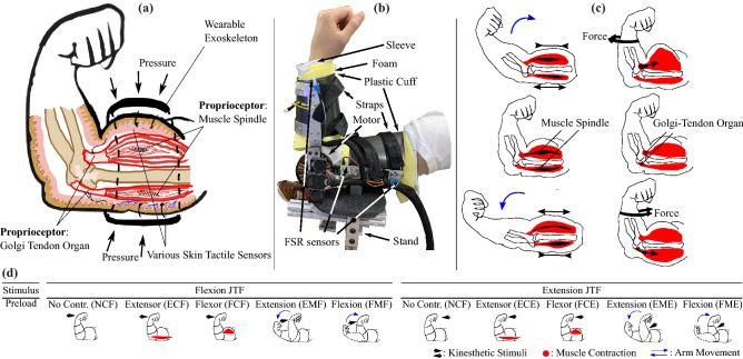
Figure 1.
(a) Illustration of dissected arm view, showing the primary proprioceptors involved in joint torque sensing as well as how the exoskeleton applies pressure and masks skin receptors. (b) Picture of a participant’s arm with the exoskeleton worn, showing the exoskeleton components. (c) Illustration of how each propioceptor is triggered under different joint conditions. The left column shows how the Muscle Spindles (MS) lengthen or shorten depending on the arm’s movement. The right column illustrates how isometrically contracted flexor or extensor activity affects the Golgi–Tendon Organ (GTO). (d) Illustration of the ten studied torque interactions, showing the kinesthetic stimuli (black arrows), contracted muscles (red highlighted muscle shapes), and direction of arm motion (blue arrows).

