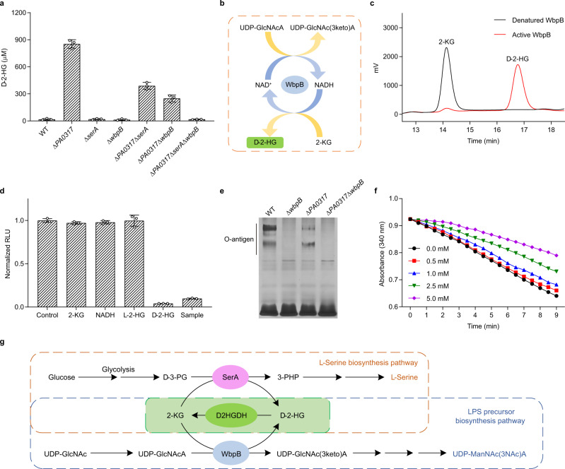
Fig. 6. Identification of the d-2-HG metabolism pathways in P. aeruginosa PAO1 using Bd2HG-1.
a Determination of extracellular d-2-HG of P. aeruginosa PAO1 and its derivatives by Bd2HG-1. The concentrations of DhdR and Bio-dhdO-1 are 0.3 and 1 nM, respectively. Data shown are mean ± s.d. (n = 3 independent experiments). b Schematic diagram of WbpB-catalyzed coupled reaction. c HPLC analysis of the product of WbpB-catalyzed 2-KG reduction. The reaction mixtures containing 2-KG (5 mM), NADH (5 mM), and active or denatured WbpB (0.4 mg mL−1) in 50 mM Tris-HCl (100 mM NaCl, pH 7.4) were incubated at 37 °C for 3 h. Black line, the reaction with denatured WbpB; red line, the reaction with active WbpB. d Chiral analysis of WbpB produced 2-HG by Bd2HG-1. The concentration of 2-KG, NADH, l-2-HG, and d-2-HG is 20 μM. The concentrations of DhdR and Bio-dhdO-1 were 0.3 and 1 nM, respectively. The sample is 50-fold diluted WbpB-catalyzed product. Data shown are mean ± s.d. (n = 3 independent experiments). e SDS-PAGE analysis of LPS of P. aeruginosa PAO1 and its derivatives. f Inhibitory of d-2-HG toward WbpB. The reaction mixtures containing WbpB (0.3 mg mL−1), 2-KG (0.5 mM), NADH (200 μM), and 0–5 mM d-2-HG in 50 mM Tris-HCl (100 mM NaCl, pH 7.4) were incubated at 37 °C for 9 min. g d-2-HG metabolism pathways in P. aeruginosa PAO1. The coupling between D2HGDH and SerA or D2HGDH and WbpB makes the robust interconversion between d-2-HG and 2-KG, and facilitates the biosynthesis of l-serine (orange dotted box) and precursor of LPS in P. aeruginosa PAO1 (blue dotted box). Source data are provided as a Source Data file.

