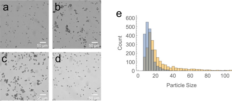
Fig. 6. A bead-based assay to measure aptamer binding to cell-surface N-glycans.
Paraformaldehyde-fixed Dictyostelium discoideum cells were incubated with (A) streptavidin beads (negative control), (B) concanavalin A (ConA)-coupled beads (positive control), (C) aptamer i-6-coupled beads, or (D) aptamer i-6-coupled beads after 1 h of preblocking with ConA. Cells were imaged with a 20X objective and scale bars correspond to 50 microns. E Histogram of particle sizes (number of pixels) for i-6 aptamer beads incubated with fixed cells either with (blue) or without (orange) ConA blocking. Images for this analysis were taken with a 5X objective (Supplementary Fig. 10). Source data are provided as a Source Data file.

