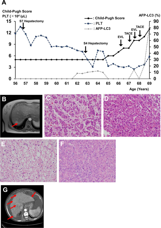
Fig. 1. Hepatocellular carcinoma (HCC) in a female patient with GSD IXc.
A Clinical course. Deterioration of cirrhosis and HCC progressed with age. B Liver MRI. T2-weighted imaging detected a 2-cm tumor in the liver (S7 section). C–F Pathological tissues. The S7 tumor exhibited characteristics of well-differentiated (C) and moderately differentiated (D) HCC combined with hepatocytes with a clear cell cytoplasm (E), consistent with pathology in GSD. The S4 tumor was poorly differentiated (F) and moderately differentiated HCC. G Liver CT. Multiple HCC and ascites were detected in the abdomen.

