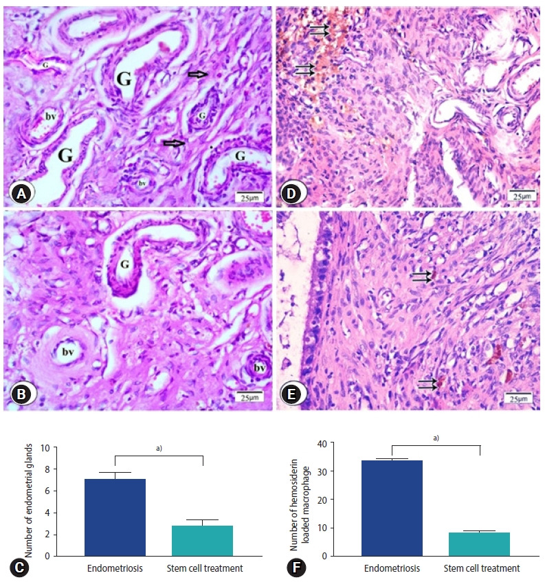
Figure 4.

Photomicrographs of endometriosis lesions from the endometriosis group (A, D) and the stem cell treated group (B, E) stained by H&E showing (A) multiple endometrial glands (G), stromal cells (arrows) and multiple blood vessels (bv) in endometriosis group (B) few endometrial glands surrounded by blood vessels. (C) Number of endometrial glands. (D) Endometriosis group; numerous hemosiderin-laden macrophages (double arrows), (E) stem cell treated group; few hemosiderin-laden macrophages were found (double arrows). (F) Number of hemosiderin loaded macrophage. (C, F) Values are presented as mean±standard error of the mean. a)p<0.001.
