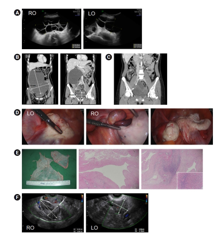
Figure 1.

(A) Sonograms of multilocular ovarian cysts (right and left) discovered early in pregnancy (12 weeks). (B) Computed tomography (CT) views of the same cysts, 10 weeks postpartum. (C) CT image showing a smaller left ovary (LO) and enlarged right ovary (RO) with twisted pedicle (arrows), 9 months postpartum. (D) Operative photos of the shrunken left ovary, the enlarged right ovary with a twisted pedicle, and the right ovary after torsion release and cyst enucleation. (E) Gross findings of the right ovary (left), with a benign denuded wall and mildly edematous stroma (H&E sections; original magnification, ×40; inset ×200). (F) Sonograms of normally appearing ovaries 10 weeks after surgery.
