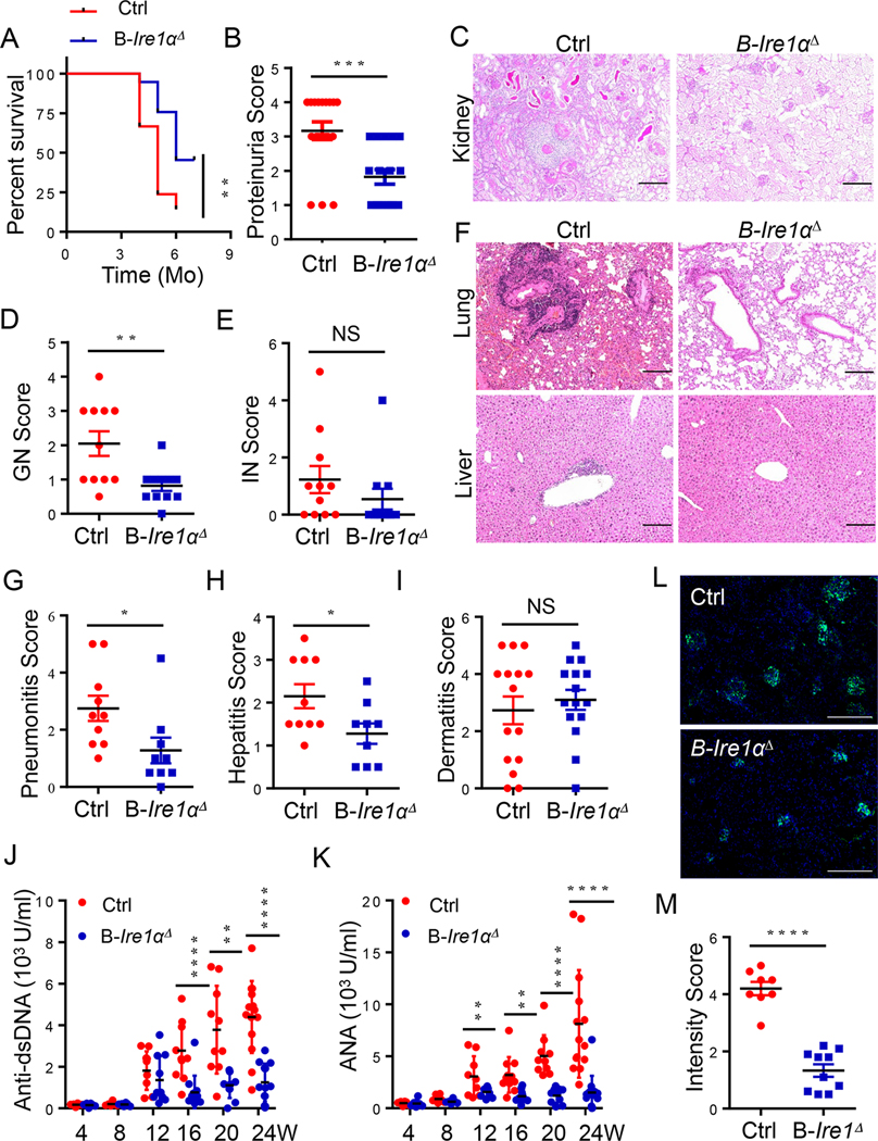
Fig. 2. B cell-intrinsic IRE1α expression is responsible for SLE progression and rapid mortality in murine lupus.
(A) Survival of Cd19-Ire1αΔ mice and their littermate control mice (n=10). (B) Proteinuria scores of 16-week-old mice. (C-E) Representative low magnification images of PAS-stained kidneys sections (C), and scores of glomerular nephritis (D) and interstitial nephritis (E) were shown (n=11;). (F) Representative images of H&E–stained lung (top) and liver sections (bottom) illustrating perivascular infiltrates in target organs from 20-week-old mice. (G-I) Pneumonitis scores (G), Hepatitis scores (H), and Dermatitis scores (I) of control and Cd19-Ire1αΔ mice. (J &K) ELISA analysis of anti-dsDNA (J) and ANA (K) in serum from Cd19- Ire1αΔ mice and their litter controls. (L & M) Representative immunofluorescence histology staining of kidney glomeruli (L) and data analysis (M) of anti-IgG (Green) were shown (Red, n=8; blue, n=10). Significance determined by Gehan-Berslow-wilcoxon test (A) and an unpaired Student’s t-test (B, D, E, G-K, & M). * p<0.05, ** p<0.01, *** p<0.001, and **** p<0.0001. Scale bars, 200 μm (C, F & L).

