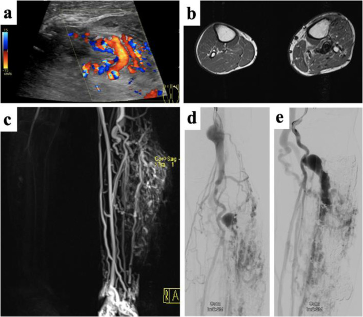
Fig. 4.
Arteriovenous malformation of the lower limb with associated soft-tissue proliferation. a Color-coded Duplex ultrasound reveals vigorous flow along the AVM nidus. b Cross-sectional MRI reveals multiple flow-voids as sign of arterialized flow patterns together with soft-tissue proliferation adjacent to the AVM. The mass was clinically pulsatile without ulceration at the time of clinical presentation. c Dynamic MR angiography shows rapid filling of the AVM along the left lower limb at time with only initial enhancement of the arteries of the non-affected leg. Pre-embolization angiogram in the late arterial phase (d) and early venous phase (e) depicting dilated aneurysms along the venous outflow tract

