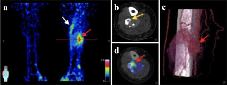
Fig. 5.
68Ga-RGB PET/CT in 38-year-old man with right-leg AVM. Maximal-intensity 68Ga-RGD PET projection (a), CTA (b) and 68Ga-RGD PET/CT (d), and 3D reconstruction of 4D-CTA (c) show nidus (SUVmax 3.2; SUVpeak 2.5, red arrows). Axial plane in (b) and (d) is at location of line in (a). Heterogeneous pattern of enhanced uptake is seen in tissue adjacent to nidus at more proximal part of the right leg (white arrow); bone deformation in the fibula caused by compression and infiltration of vessels is also seen (yellow arrow), along with large venous aneurysm (asterisk). Arterial flow with nidus and fistula is seen in (c), along with upcoming venous flow and the large venous aneurysm. This research was originally published in JNM. Lobeek D, Bouwman FCM, Aarntzen EHJG, Molkenboer-Kuenen JDM, Flucke UE, Nguyen HL, Vikkula M, Boon LM, Klein W, Laverman P, Oyen WJG, Boerman OC, Terry SYA, Schultze Kool LJ, Rijpkema M. A Clinical Feasibility Study to Image Angiogenesis in Patients with Arteriovenous Malformations Using 68Ga-RGD PET/CT. J Nucl Med 2020; 61:270–275.© SNMMI

