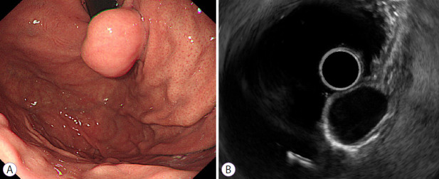
Fig. 3.
A leiomyoma in a 40-year-old man. (A) Endoscope image showing an elongated subepithelial tumor without a mucosal ulcer in the gastric cardia. (B) Endoscopic ultrasonogram revealing a homogenous hypoechoic mass without anechoic spaces or hyperechoic spots arising from the fourth layer measuring 2.1×1.5 cm in size. According to the nomogram, the sum of values is 22, corresponding to a risk probability of 0.204. This result indicates a high probability of a leiomyoma.

