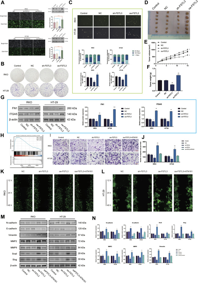
FIGURE 4.
FSTL3 overexpression promotes a malignant phenotype in colorectal cancer (CRC). (A) The transfection efficiency was verified by the GFP expression as well as the western blot. Transfection efficiency is represented as percentages. (B) Clone formation capacity of the CRC cells transfected with the NC, sh- FSTL3, and oe-FSTL3 constructs assessed using the clone formation assay. (C) Representative images from the sphere-forming assay. The number of tumorspheres was counted and plotted, and percentage of tumorspheres with diameters of 50–100 μm, 100–150 μm, or >150 μm was calculated and plotted. The scale bar represents 100 μm. (D) The xenograft tumors from the nude mice. (D–F) (E) Tumor volume and (F) weight of xenografts from the nude mice. (G) FN1, ITGA5 levels in CRC cells transfected with NC, sh-FSTL3, and oe-FSTL3 were examined using western blot. (H) Gene Set Enrichment Analysis (GSEA) of FSTL3. (I,J) The invasion ability of CRC cells after transfection; the relative invasive cell number is shown towards the right. (K,L) The migratory ability of the different groups of CRC cells [(K): RKO, (L): HT-29] was examined using the wound-healing assays. (M,N) Expression of the EMT-related proteins was examined using western blots after the transfection of the CRC cells with NC, sh- FSTL3, and oe-FSTL3 constructs and treatment with 10 μM ATN-161, an antagonist of integrin α5β1. The statistical analysis of the western blot result is shown towards the right. * p < 0.05, ** p < 0.01, *** p < 0.001.

